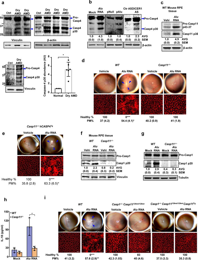
Figure 1. Caspase-4/11 in geographic atrophy and RPE degeneration.

(a) Left and top quadrants, immunoblots for pro-caspase-4 (pro-Casp4) and the p30 cleavage product of caspase-4 (Casp4 p30) in the RPE of human eyes with geographic atrophy (dry AMD) as compared to unaffected controls (Ctr). Specific bands of interest are indicated by arrowheads. Lower right quadrant, densitometry of the bands corresponding to caspase-4 p30 normalized to loading control. The molecular weight markers are indicated on the left side of the blot (Data are presented as mean ± SD; n = 3 control eyes; n = 6 dry AMD eyes; *P = 0.002, two-tailed t test). (b) Immunoblots for pro-Casp4 and Casp4 p30 in human RPE cells mock transfected (just transfection mixture) or transfected with Alu RNA; Alu expression plasmid (pAlu) or empty vector (pNull); or DICER1 or control (Ctr) anti-sense oligonucleotides (AS). Specific bands of interest are indicated by arrowheads. (c) Immunoblot for pro-caspase-11 (Pro-Casp11) and the p30 cleavage product of caspase-11 (Casp11 p30) in RPE tissue of WT mice injected subretinally with Alu RNA or vehicle (Vehi). n = 3 mice per group. (d,e) Top, fundus photographs of the retinas of WT (n = 8 eyes) and Casp11−/− (n = 10 eyes) mice, (d) and Casp11−/− (n = 8 eyes) mice expressing a human caspase-4 transgene (Casp11−/− hCasp4Tg) (e) injected with vehicle or Alu RNA. The degenerated retinal area is outlined by blue arrowheads. Bottom, immunostaining with zonula occludens-1 (ZO-1) antibody to visualize RPE cellular boundaries; loss of regular hexagonal cellular boundaries is indicative of degenerated RPE. (f) Immunoblots of pro-caspase-1 (pro-Casp1) and the p20 cleavage product of caspase-1 (Casp1 p20) in RPE tissue of WT and Casp11−/− mice injected subretinally with vehicle (Vehi) or Alu RNA. n = 3 mice per group. (g) Immunoblots of pro-caspase-1 and the p20 cleavage product of caspase-1 in WT and Casp11−/− mouse RPE cells treated with Alu RNA. (h) IL-18 secretion by WT and Casp11−/− mouse RPE cells mock transfected or transfected with Alu RNA. n = 3 independent experiments. Data presented are mean ± SD; *P = 0.014, two-tailed t test. (i) Top, fundus photographs of the retinas of WT (n = 8 eyes), caspase-1 and caspase-11 deficient (n = 7 eyes) mice (Casp1−/− Casp11129mt/129mt) as well as Casp1−/− Casp11129mt/129mt (n = 8 eyes) mice expressing functional mouse caspase-11 from a bacterial artificial chromosome transgene (Casp1−/− Casp11129mt/129mt Casp11Tg) subretinally injected with vehicle or Alu RNA. For all immunoblots, cropped gel image of bands of interest of representative immunoblots of three independent experiments and densitometric analysis (mean (SEM)) are shown. Tubulin or β-actin or Vinculin was as a loading control as indicated in each blot. In d, e and i, binary (Healthy %) and morphometric quantification (PM, polymegethism (mean (SEM))) of RPE degeneration are shown (Fisher’s exact test for binary; two-tailed t test for morphometry; *P < 0.05; **P < 0.01; ***P < 0.001). The degenerated retinal area is outlined by blue arrowheads in the fundus images. Loss of regular hexagonal cellular boundaries in ZO-1 stained flat mounts is indicative of degenerated RPE.
