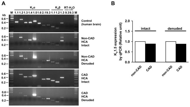
Figure 1. The mRNA expression of KV1 channel subunits in human coronary arteries.
Representative gel images of RT-PCR amplification products from one endothelium-intact and one endothelium-denuded from non-CAD and CAD HCAs are shown in figure 1A. KVα1.2, 1.3, 1.5, 2.1, 9.3 and KVβ1.1–1.3 were consistently found in different arterial samples, whereas KVα1.1 1.4 and 1.6 subunits were variably detected. As a positive control, human brain tissue from a normal subject was found to express all KV channel subunits studied (top). RT-, without reverse transcription; H2O, without template; M, marker. Quantitative PCR revealed a slight reduction of KVα1.5 mRNA in CAD arteries (B).

