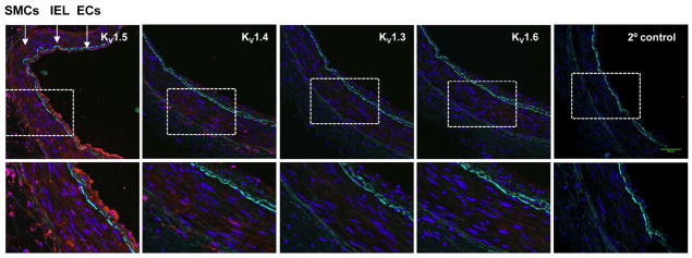
Figure 3. Immunofluorescence localization of KV1 α-subunits in human coronary arteries.
Representative confocal images show KV1 α-subunit proteins (red) in cross tissue sections (10 μm) of an intact non-CAD HCA. Amplified regions indicated by dotted outline are shown in bottom panels. KV1.5 was highly expressed in both smooth muscle cells (SMCs) and endothelial cells (ECs), with a modest expression of KV1.4 in SMCs. KV1.3 and KV1.6 were faintly detected in SMCs and/or ECs. Cell nuclei were stained with DAPI (blue). IEL, internal elastic lamina (IEL, green autofluorescence).

