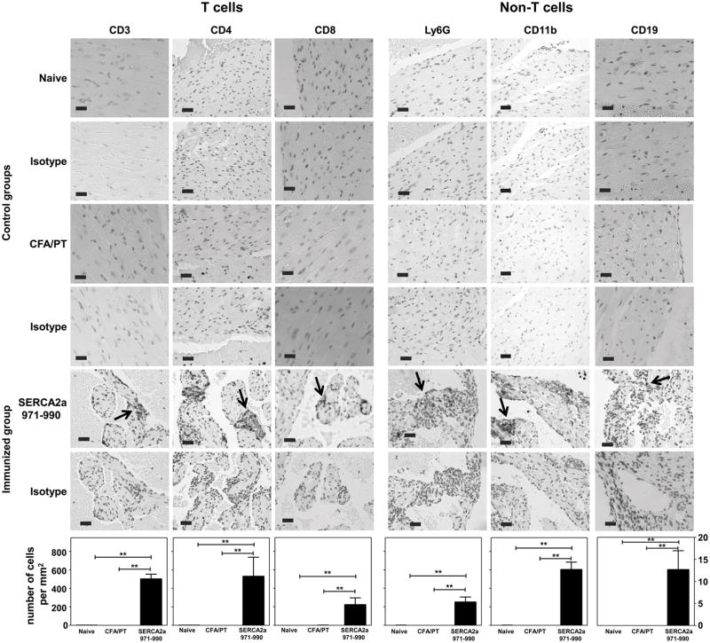
Figure 2. Inflammatory infiltrates in hearts from animals immunized with SERCA2a 971-990 reveals the presence of T cells and non-T cells.
Heart sections derived from mice immunized with or without SERCA2a 971-990 were evaluated for the presence of T cells (CD3, CD4 and CD8) and non-T cells (Ly6G, CD11b and CD19) by immunohistochemistry. Ag retrieval was performed on the deparaffinized sections as described in the methods section. Numbers of immunopositive cells were then determined using the nuclear V9 software. Each bar represents mean ± SEM values (n= 4 to 6 mice per group). **p ≤ 0.01.

