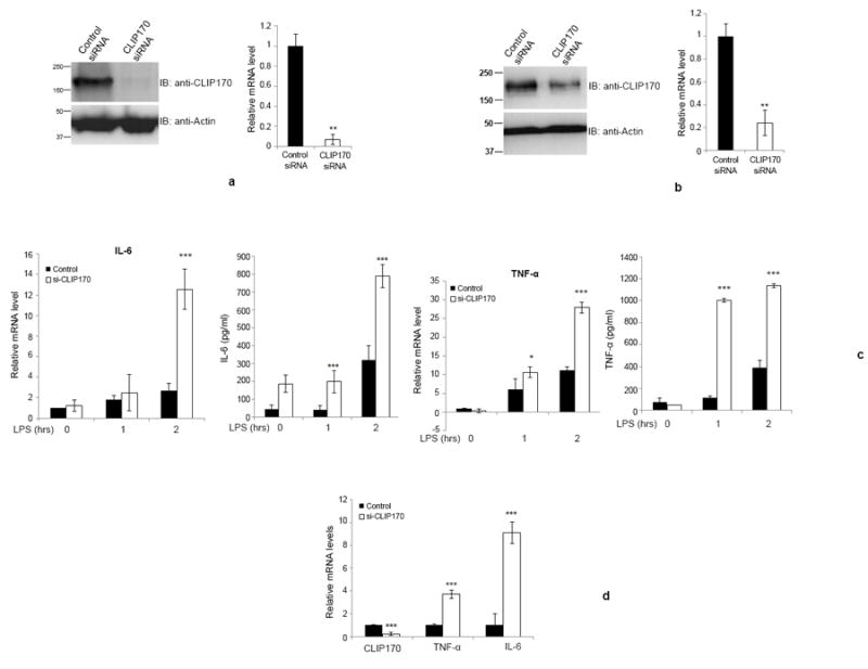
Figure 5. Silencing of CLIP170 enhances LPS-induced pro-inflammatory cytokines in mice.

(a & b) Depletion of CLIP170 in splenocytes or Kupffer cells derived from CLIP170 siRNA treated mice. Mice were treated with CLIP170 siRNA or control siRNA for 48 h, followed by collection of splenocytes (a) or Kupffer cells (b). The cells were analyzed by immunoblotting and qPCR to examine the levels of endogenous CLIP170. The blot was probed with anti-CLIP170 to detect the endogenous CLIP170. Actin served as the loading control. (c) Depletion of CLIP170 potentiates the expression of IL-6 and TNF-α in splenocytes. The splenocytes isolated from mice injected with CLIP170-specific or control siRNA were challenged with LPS (100 ng/ml) for the indicated time points followed by quantification of IL-6 and TNF-α by ELISA and qPCR. (d) Potentiated levels of IL-6 and TNF-α in the liver of CLIP170-silenced mice. Seventy-two hours after the siRNA delivery, mice were injected with LPS (25 mg/kg) intraperitoneally and the levels of IL-6 and TNF-α were quantified by qPCR. Data are presented as mean ± SD from two independent experiments of six mice per group; *P < 0.05, **P < 0.01 and ***P < 0.001.
