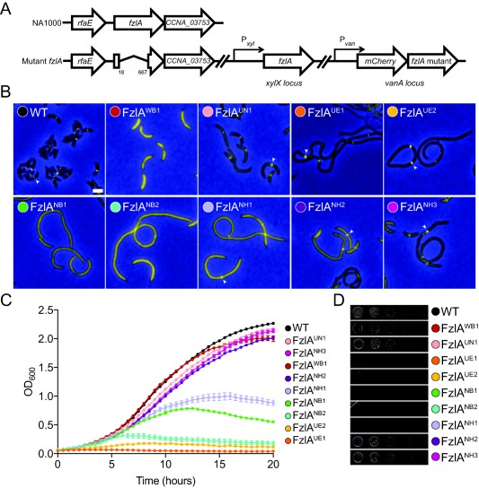
Figure 3.

fzlA mutant strains display a range of division and localization deficiencies.
A. Cartoon depicting genetic backgrounds for in vivo testing. In these strains, fzlA has been deleted at the native locus and a xylose‐inducible copy of fzlA is at the xylX locus. A vanillate‐inducible copy of the mCherry‐fzlA variant is integrated at the vanA locus.
B. Merged fluorescence and phase contrast microscopy images depicting mCherry‐FzlA mutant protein (yellow) localization in cells depleted of WT FzlA and grown with vanillate to induce the indicated mCherry‐FzlA variant for 24 h prior to imaging. White arrowheads mark localized FzlA bands. Scale bar = 2 μm.
C. Growth curves of the same strains as in (B) grown with vanillate and depleted of WT FzlA for 24 h prior to the start of the experiment. Mean of three technical replicates ± SEM is shown.
D. Spot dilutions of strains as in (B), plated on PYE agar with vanillate, without predepletion of WT FzlA. Strain key: WT (EG1310), FzlAWB1 (EG1435), FzlAUN1 (EG1312), FzlAUE1 (EG1313), FzlAUE2 (EG1621), FzlANB1 (EG1430), FzlANB2 (EG1438), FzlANH1 (EG1311), FzlANH2 (EG1441), FzlANH3 (EG1442).
