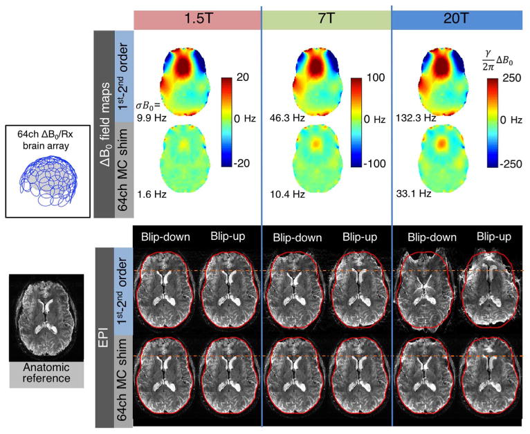
Fig. 13.

ΔB0 field maps and simulated EPI distortion for 1.5 T, 7 T, and 20 T brain imaging. The 1.5 T and 20 T ΔB0 field maps are scaled versions of an acquired 7 T brain field map. Two cases are shown: global 1st–2nd order SH shimming and global 1st–2nd order SH shimming+slice-optimized 64ch MC shimming using the close-fitting ΔB0/Rx helmet coil configuration shown in Fig. 11. Geometric distortion is applied to a T2*-weighted anatomic reference image using FSL FUGUE for an echo spacing of 0.56 ms with GRAPPA acceleration of R = 2 (effective echo spacing of 0.28). Both anterior-posterior and posterior-anterior distortions are simulated to emphasize the distortion. ΔB0 and EPI voxel shifts scale linearly with field strength. While negligible at 1.5 T, the distortion becomes pronounced at 7 T, and at 20 T the images are cartoonishly warped. The voxel shifts are 13 times larger at 20 T than at 1.5 T. The 64ch multi-coil shim array is capable of reducing the distortion to less than what occurs at 7 T with global 2nd order shimming (σB0 within the slice is 33.1 Hz for 20 T MC shim versus 46.3 Hz for 7 T 1st–2nd order SH shim). This highlights the need for high-spatial order shimming systems to enable functional MR imaging on envisioned scanners of the future with field strengths well above 7 T.
