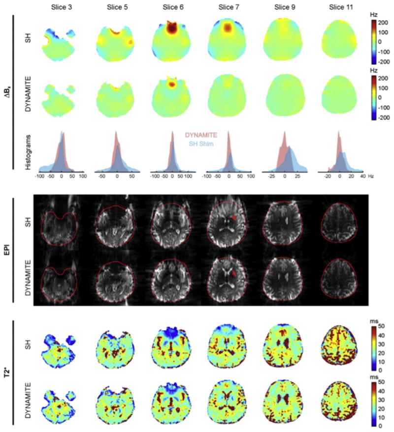
Fig. 8.

Reproduced with permission from Juchem et al. (2011). Data from 6 brain slices comparing experimental 1st–3rd order global SH shimming with 48ch single slice optimized dynamic MC shimming using the array shown in Fig. 6b. For each of the two shim methods, ΔB0 field maps, voxel ΔB0 histograms, multi-shot EPI images, and T2* maps are shown. Global SH shimming removes smoothly-varying components of B0, but provides limited improvement in areas with steep B0 variation such as the frontal lobes. Dynamic MC shimming helps mitigates these areas of peak ΔB0 and reduces geometric distortion both inside the brain (red arrow) and at the brain surface. Signal voids caused by through-slice dephasing are also mitigated. After MC shimming, the T2* distributions more closely match the values expected for the underlying tissue types (especially in the frontal lobe region of slice 7). The MC shim currents were updated in 1.5 ms and no artifacts were observed due to eddy currents or other transient effects. Across 5 human subjects, the average reported σB0Global values were 32.3 Hz for the global 1st–3rd order shims and 13.3 Hz for the dynamic MC shims, a 59% difference.
