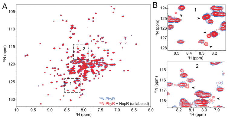
Figure 4. NepR induces structural shifts in unphosphorylated PhyR.
(A) 1H-15N TROSY spectra of 15N-labeled C. crescentus PhyR (blue) overlaid with 15N-PhyR in the presence of 1.25-fold molar excess of unlabelled C. crescentus NepR (red). Boxes 1 and 2 are magnified in panel B. (B) Selected PhyR peak shifts observed in the presence of NepR are marked with black arrowheads.

