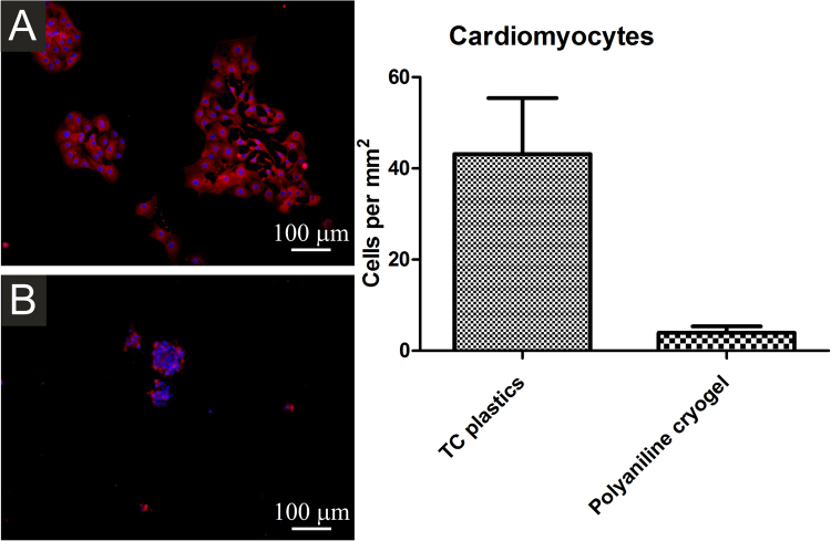
Figure 9.
Isolated cardiomyocytes seeded onto TC plastics (A) and polyaniline cryogel (B). Cardiomyocytes were visualised using antibody against cardiomyocyte-specific myosine heavy chain (red). Individual cells were visualised through nuclei counterstaining by DAPI (blue). Number of cells at the starting point is not presented as the cardiomyocytes did not proliferate after the seeding. The number of cardiomyocytes adhered on the cryogel was significantly smaller than on TC plastic. The micrographs were taken on day 2 after seeding.

