Figure 4.
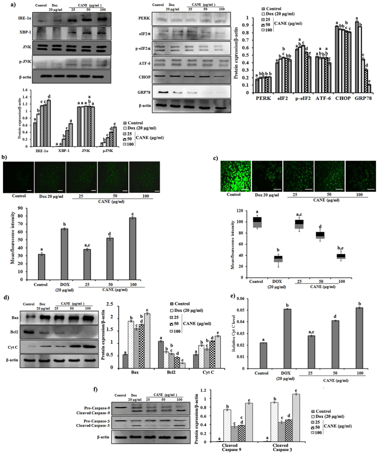
CANE sensitizes A549 cells to follow ER stress leads to mitochondria-mediated apoptotic pathway. (a) Western blotting of ER stress markers, β-actin used as loading control. Docetaxel (20 μg/ml) was used as a positive control while the surfactant-polysorbate 80 mix devoid of carvacrol was also used as a control. Densitometry analysis of IRE1-α, PERK, and ATF-6 was determined by Image J software. (b) Evaluation of intracellular Ca+2 accumulation. (c) Mitochondrial membrane potential of A549 cells treated with CANE (25-100 μg/ml). Fluorescent images were captured at 20X magnification [scale bar = 0.1 mm]. Image J software was used to determine the mean fluorescence intensity. (d) Effect of CANE on expression of Bax, Bcl2, and Cyt C in A549 cells. (e) Cyt C level in CANE-treated cells measured by spectrophotometric method. (f) Effect of CANE on expression of cleaved caspase-9, and 3 in A549 cells. Each value in the graph represents as the mean ± SD of three independent experiments. Values with different superscripts differ significantly from each other (p < 0.05).

