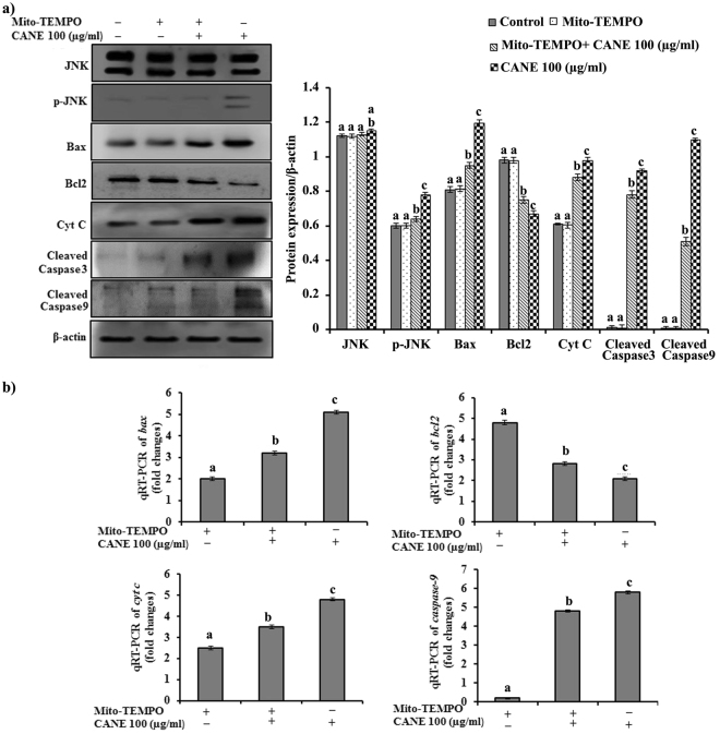
Figure 6.
Effect of ROS inhibition using Mito-TEMPO on the vital apoptotic markers at translational and transcriptional level. (a) Cellular levels of protein markers JNK, p-JNK, Bax, Bcl2, Cyt C, caspase-3, caspase-9, and β- actin in A549 cells incubated with Mito-TEMPO (10 μM). Densitometry analysis of the respective proteins was evaluated by Image J software, and results were normalized with β-actin with respect to controls. (b) Quantitative RT-PCR (qRT-PCR) analysis of bax, bcl2, cyt c, caspase-9, and β-actin after treatment with CANE in conjunction with Mito-TEMPO. Mito-TEMPO controls expression of apoptotic genes at transcription level represented in fold change compared with control. Each value in the graphs represents as the mean ± SD of three independent experiments. Values with different superscripts differ significantly from each other (p < 0.05).

