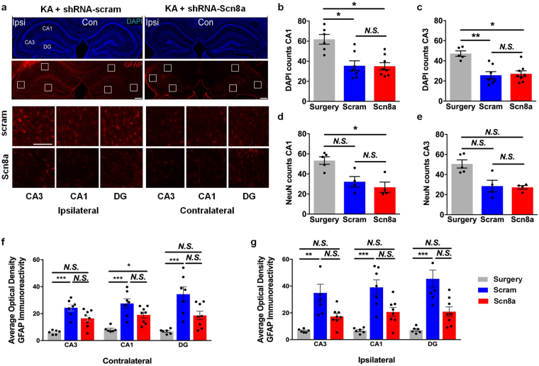
Figure 5.
Scn8a knockdown reduces GFAP expression in a mouse model of MTLE. (a) Representative images of hippocampi following kainic acid (KA) and either control shRNA-scram (left column) or shRNA-Scn8a (right column) injections. DAPI staining (top) shows the hippocampal ultrastructure. GFAP immunoreactivity (bottom) was used to compare the extent of reactive gliosis in AAV-treated MTLE mice. Scale bar, 300 µm. Inset images show CA3, CA1, and DG regions of the ipsilateral (left panel) and contralateral (right panel) hippocampi following treatment with either shRNA-scram (top row) or shRNA-Scn8a (bottom row). Scale bars, 100 µm. Approximately 50% reduction in number of cells (b,c) and neurons (d,e) in the ipsilateral hippocampus were observed in MTLE animals treated with either shRNA-scram (blue) or shRNA-Scn8a (red) compared to surgery-only controls (gray). (f,g) Quantification of optical density (O.D.) of normalized GFAP immunoreactivity in the contralateral and ipsilateral hippocampi in surgery-only (gray) or MTLE animals treated with either shRNA-scram (blue) or shRNA-Scn8a (red). GFAP expression was comparable between the surgery-only controls (gray) and shRNA-Scn8a-treated MTLE mice (red) in the ipsilateral hippocampus. In contrast, GFAP expression was significantly increased in the shRNA-scram-treated MTLE mice compared to surgery-only controls in both the ipsilateral and contralateral hippocampus. Non-parametric Kruskal-Wallis test followed by Dunn’s multiple comparisons test (NeuN: N = 4–5/group, DAPI and GFAP: N = 6–8/group). All data are presented as mean ± SEM. *p < 0.05, **p < 0.01, ***p < 0.001, N.S, not significant.

