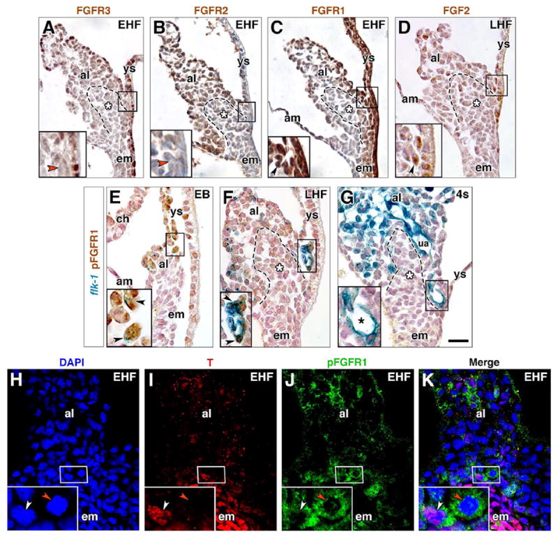
Fig. 4. Localization of FGF members within the nascent VOC.

(A-G) Immunostaining for FGFR3 (A), FGFR2 (B), FGFR1 (C), FGF2 (D), all at the headfold stages, and pFGFR1 (E-G) in EB through 4s stage flk-1: lacZ reporter specimens. Boxed region in main panels is the prospective VOC (A-E) or endothelialized VOC (F, G) region, enlarged in insets, lower left. Dashed outline, posteriormost (distalmost) extension of the primitive streak, and white asterisk, ACD (A-D, F, G). Red arrowheads (A, B), FGFR-3- and FGFR-2-negative cells. Black arrowhead (C), FGFR1-positive rosette. Black arrowhead (D), FGF2-positive cell. Black arrowheads (E, F), flk-1-positive/pFGFR1-positive angioblasts. Black asterisk (G), flk-1-positive/pFGFR1-negative mature VOC. (H-K) EHF stage, frontal (ventral) optical section through ventral allantoic and embryonic tissue stained with DAPI (H), T (I), pFGFR1 (J); channels merged (K). Insets are enlargements of prospective VOC, boxed in main panels. White arrowhead, T-positive/pFGFR1-positive angioblast; red arrowhead, pFGFR1-positive/T-negative angioblast. Scale bar (G): 20 μm (A-K). al, allantois; am, amnion; ch, chorion; em, embryo; ua, umbilical artery; ys, yolk sac.
