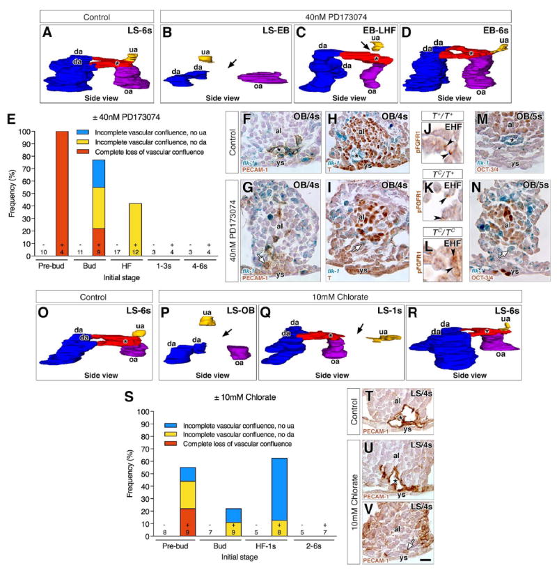
Fig. 5. Stage-specific failure of formation of the vascular confluence in PD173074- or chlorate- treated specimens.

“Control” indicates untreated conceptuses that were cultured alongside pharmacologically-treated ones until 7–8s stage. Transverse histological sections (F-I, M, N, T-V) are oriented with ventral toward the bottom. Black asterisk indicates the VOC (A, C, D, F, H, M, O, Q, R, T, U). (A-D, O-R) 3D models, in side view (posterior to the right), reconstructed from post-culture PECAM-1-immunostained arterial vessels at the posterior embryonic-extraembryonic interface of control (A, O), PD173074-treated (B-D) or chlorate-treated (P-R) conceptuses (initial stages, upper right). Color key as in Fig. 2. Black arrows indicate missing arterial connections due to loss of the VOC (B, P) or to incomplete branching of the VOC (C, Q). (E, S) Frequency of anomalies observed in the vascular confluence in post-culture specimens from PD173074 (E) or chlorate (S) experiments by their initial stage, sample size and group’s treatment (± PD173074 or chlorate). (F, G) Transverse sections in the flk-1: lacZ reporter showing the PECAM-1-positive endothelialized VOC in the control (F), but non-endothelialized angioblasts in the prospective VOC of PD173074-treated specimens (G, white arrow). (H, I) Transverse sections in the flk-1: lacZ reporter showing down-regulation of T in the endothelialized control VOC (H) and in nonendothelialized angioblasts of the prospective VOC in PD173074-treated specimens (white arrow) (I). (J-L) Histological sections through the prospective VOC showing that pFGFR1 is unaffected in the three TC genotypes. (M, N) Transverse sections in the flk-1: lacZ reporter showing down-regulation of OCT-3/4 in the endothelialized VOC of untreated controls (M) and in non-endothelialized angioblasts of the prospective VOC in PD173074-treated specimens (N, white arrow). (T-V) Transverse histological sections showing the PECAM-1-positive endothelialized VOC in a control (T) and a chlorate-treated specimen (U), and non-endothelialized angioblasts in the prospective VOC of another chlorate-treated specimen (V, white arrow). Scale bar (V): 6 μm (J-L); 25 μm (F-I, M, N, T-V). al, allantois; da, dorsal aortae; omphalomesenteric artery, oa; umbilical artery; ys, yolk sac.
