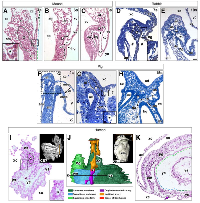
Fig. 6. Conserved features at the posterior embryonic-extraembryonic interface of placental mammals.

For all panels: black asterisk (A-E, G-I) indicates VOC; white asterisk (A-E, G-I) and dashed outline (A, D, G, I) indicate dense allantoic core; black dashed line (B, C, H) indicates arterial vessels out of sectional plane; colored dashed lines (A, K) indicate distinct region of visceral endoderm with color key below J; and hash mark (D, F, G) indicates artifactual spaces produced by fixation. (A-C) Posterior embryonicextraembryonic interface of a mouse conceptus at the 4s (A), 6s (B) and 8s (C) stage. VOC in A is boxed and enlarged in inset. (D, E) Posterior embryonic-extraembryonic interface of a rabbit conceptus at the 7s (D) and 10s (E) stages. VOC in D is boxed and enlarged in inset. (F-H) Posterior embryonic-extraembryonic interface of a pig conceptus at the 4s (F, G) and 15s (H) stages. (G) Enlargement of hashed region in F; VOC is boxed in main panel and enlarged in inset. (I-K) Posterior embryonic-extraembryonic interface of a human conceptus at Carnegie stages 7 (I) and 8 (J,K). Volume-rendered models (insets I, J) with transverse sections (I, K) oriented ventral toward the bottom and taken at the level of vertical line (inset I) and horizontal line (J). 3D model (J, color key below), reconstructed from nascent arterial vessels and endoderm (boxed region, inset J). VOC in I is boxed and enlarged in inset. Purple arrow (K) indicates omphalomesenteric artery (oa). Scale bar (E): 20 μm (A, B, I); 40 μm (C, D, G); 90 μm (F); 100 μm (E, H); 300 μm (Insets I, J); 400 μm (K). ac, amniotic cavity; ad, allantoic diverticulum; am, amnion; al, allantois; cs, connecting stalk; da, dorsal aorta; em, embryo; endo, endoderm; hg, hindgut; meso, mesoderm; pg, primitive groove; ua, umbilical artery; xc, exocoelomic cavity; yc, yolk cavity; ys, yolk sac.
