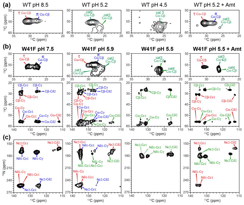
Figure 4.
H37 chemical shifts in WT (a) and W41F (b, c) AM2-TM from 2D 13C-13C (a, b) and 15N-13C (c) correlation spectra as a function of pH. (a) H37 Cα-Cβ regions of the 2D 13C-13C spectra of the WT peptide. (b) H37 Cα-Cβ and aliphatic-aromatic regions of the 2D 13C-13C spectra of the W41F mutant. (c) Aromatic region of the 2D 15N-13C correlation spectra of the W41F mutant. The pH and drug binding state of the samples are indicated. The mutant channel shows higher π tautomer intensities at high pH and more cationic histidine peaks at low pH compared to the WT channel.

