
Fig. 2.
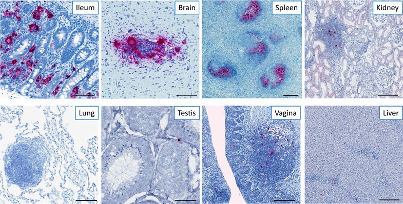
Flexibility and reproducibility of the RNAscope approach in different tissue types. Representative images of SIV RNAscope performed on chronically infected rhesus macaques in ileum, brain, spleen, kidney, lung, testis, vagina, and liver showing productively infected vRNA+ cells and viral particles on FDCs within the BCFs. Scale bars 200 μm