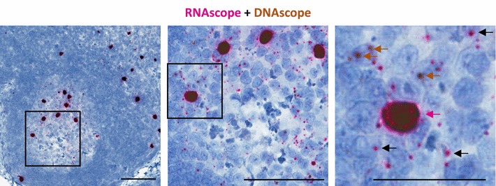
Fig. 3.

Multiplexing RNAscope and DNAscope on the same tissue section. Representative images with progressive magnification of multiplexed RNAscope and DNAscope on the same lymph node section from an SIV acutely infected rhesus macaque. In the final pane, the red arrow highlights a vRNA+/vDNA+ productively infected cell, the brown arrows highlight vRNA−/vDNA+ infected cells, and the black arrows highlight examples of the numerous SIV viral particles trapped on the FDC network. Scale bars 200 μm
