
Figure 2(A-F).
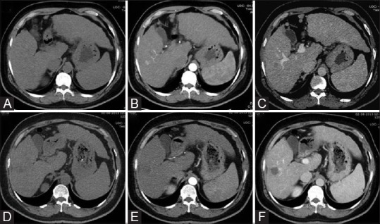
Axial triple phase CT scan images [unenhanced (A), arterial (B), and delayed (C)] shows an intraparenchymal nodule with imaging features consistent with HCC, i.e., arterial enhancement with washout. Post-RFA follow-up triple phase CT scan images 1 month after ablation [unenhanced (D), arterial (E), and delayed (F)] shows no enhancement in treated nodule, most appreciable on venous phase suggestive of complete response