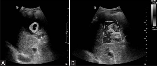
Figure 1(A and B).

Grayscale (A) and color Doppler (B) images of the gallbladder demonstrate a 3.2 cm cystic artery pseudoaneurysm within the gallbladder. On color Doppler imaging, blood flow is seen within the pseudoaneurysm in the classic “yin-yang” pattern
