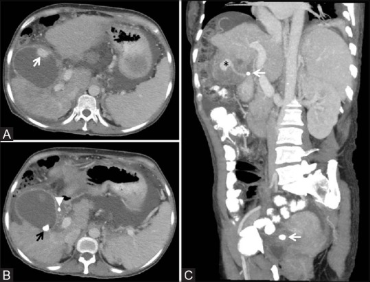
Figure 2(A-C).

Axial images from a contrast-enhanced CT (A and B) demonstrates the enhancing cystic artery pseudoaneurysm in the anterior wall of the gallbladder (A, white arrow). There are intraluminal (B, black arrow) and extraluminal (B, white arrowhead) gallstones. A coronal oblique 4 mm maximum intensity projection image (C) demonstrates the pseudoaneurysm (asterisk) being supplied by the cystic artery. Note the gallstones adjacent to the gallbladder and in the pelvis (white arrows), which were previously seen within the gallbladder on a CT from 3 months prior
