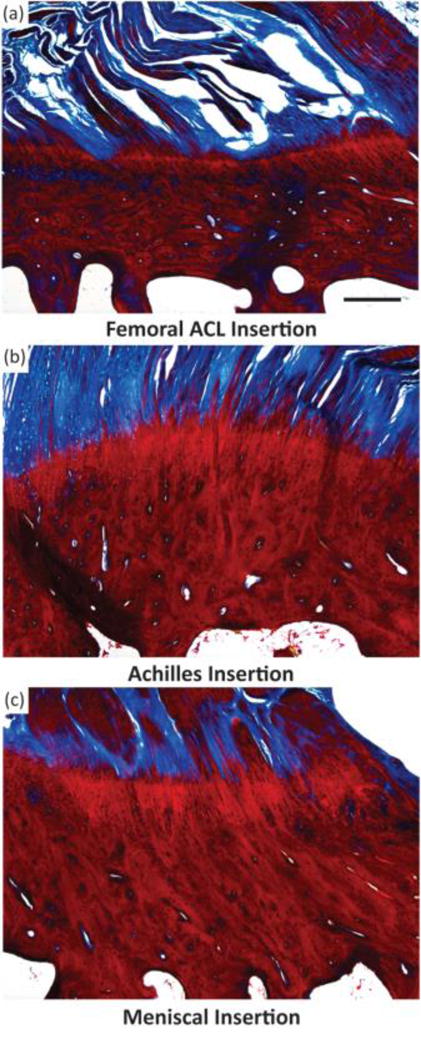
Fig. 3.

Light microscope images of three different osteochondral interfacial tissues, stained with tetrachrome stain. All images show ovine tissue, cut in the sagittal plane of the enthesis: (A) the femoral anterior cruciate ligament (ACL) insertion, (B) insertion point of gastrocnemius tendon with the calcaneal bone, referred to here as the Achilles insertion, and (C) the meniscal insertion. Trabecular pores are visible on the bottoms of each image, beneath dense calcified bone (deep red). Porous regions transition through to fibers (blue). Note the varying thicknesses of the interfacial regions, and variable morphology of the intermediate bony regions per anatomy. Scale bar is 400 μm.
