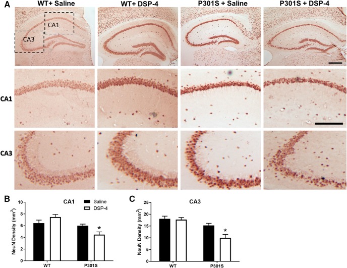
Figure 10.
LC lesions promote neurodegeneration in the hippocampus of 10 month P301S mice. Representative images (A) and quantification of NeuN IR in the (B) CA1 and (C) CA3 regions of the hippocampus of WT and P301S mice treated with saline or DSP-4. Two-way ANOVA (treatment × genotype) with Sidak's post hoc tests. Data are mean ± SEM; n = 8–12 per group. Scale bars: Top, 200 μm; Bottom, 100 μm. *p < 0.05, compared with P301S + saline.

