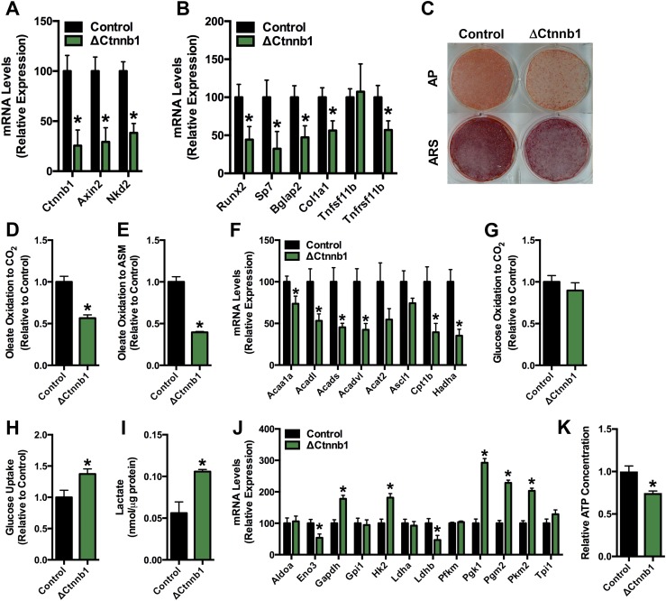
Figure 2.
Fatty acid oxidation is reduced in osteoblasts lacking β-catenin. (A) qPCR analysis of Ctnnb1, Axin2, and Nkd2 mRNA levels in primary osteoblasts isolated from Ctnnb1flox/flox (Control) and Oc-CreERT2; Ctnnb1flox/flox (ΔCtnnb1) mice treated with 1 μM tamoxifen on days 5 to 7 and then cultured in osteogenic medium for an additional 2 days. (B) qPCR analysis of markers of osteoblastic cells in control and ΔCtnnb1 osteoblasts. (C) Alkaline phosphatase (AP) and Alizarin red S (ARS) staining of cultures of control and ΔCtnnb1 osteoblasts cultured for an additional 7 days after TM administration as in (A). (D and E) Relative oxidation of 14C-Oleate to (D) 14CO2 and (E) acid-soluble metabolites (ASM) by control and ΔCtnnb1 osteoblasts. (F) qPCR analysis of genes involved in fatty acid catabolism by control and ΔCtnnb1 osteoblasts. (G) Relative oxidation of 14C-glucose to 14CO2. Results were normalized to protein concentration and then expressed relative to the control osteoblast cultures. (H) Relative uptake of 2-deoxy-D-[3H]-glucose. (I) Cellular lactate levels in control and ΔCtnnb1 osteoblasts cultured in osteogenic media for 7 days. (J) qPCR analysis of genes involved in glucose catabolism in control and ΔCtnnb1 osteoblast cultures. (K) Cellular ATP levels in control and ΔCtnnb1 osteoblasts. Results were normalized to protein concentration and then expressed relative to the control osteoblast cultures. All in vitro data were replicated in two independent studies. Data are expressed as mean ± standard error of the mean. *P < 0.05.

