Figure 6.
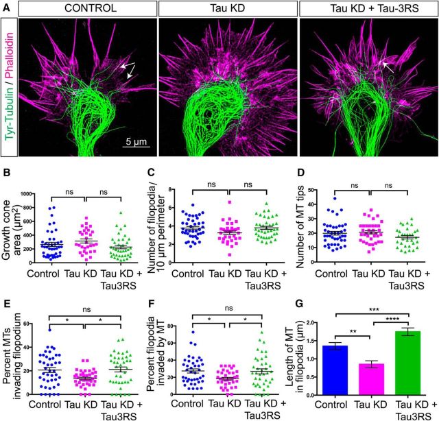
Tau knockdown prevents MT protrusion into the growth cone periphery. To obtain images of dynamic MTs in relation to actin we labeled fixed growth cones with an antibody to tyrosinated tubulin and with phalloidin, respectively, and imaged growth cones with STED microscopy (A). In a representative control growth cone MTs form a prominent loop in the central region from which individual MTs extend into the transition region. MTs reorient to enter filopodia in apposition to actin filament bundles, as shown by overlap of tubulin (green) and actin (magenta) labeling in filopodia (arrows). Following tau knockdown the MT loop becomes disorganized and individual MTs (green) only penetrate partially into the growth cone transition region containing actin labeling (magenta). These MTs fail to enter filopodia or align with actin filament bundles. Rescue experiments with human tau expression show restoration of MT bundling, MT penetration into the growth cone peripheral domain, and close MT apposition (arrow) to actin filaments in filopodia. Growth cone area (B), density of filopodia in the peripheral domain (C), and MT tips in the transition region (D) are similar between tau knockdown and control growth cones. In tau knockdown growth cones a smaller percentage of MTs in the transition region invaded the peripheral domain (E). Quantification (F, G) of filopodia invaded by MTs showed that after tau knockdown fewer filopodia contained MTs and these MTs were significantly shorter than those entering filopodia in control growth cones. Rescue with human tau restored numbers and lengths of MTs entering filopodia to control levels. ns - not significant, *p < 0.05, **p < 0.01, ***p < 0.001, ****p < 0.0001. Scale bar, 5 μm.

