
Figure 4.
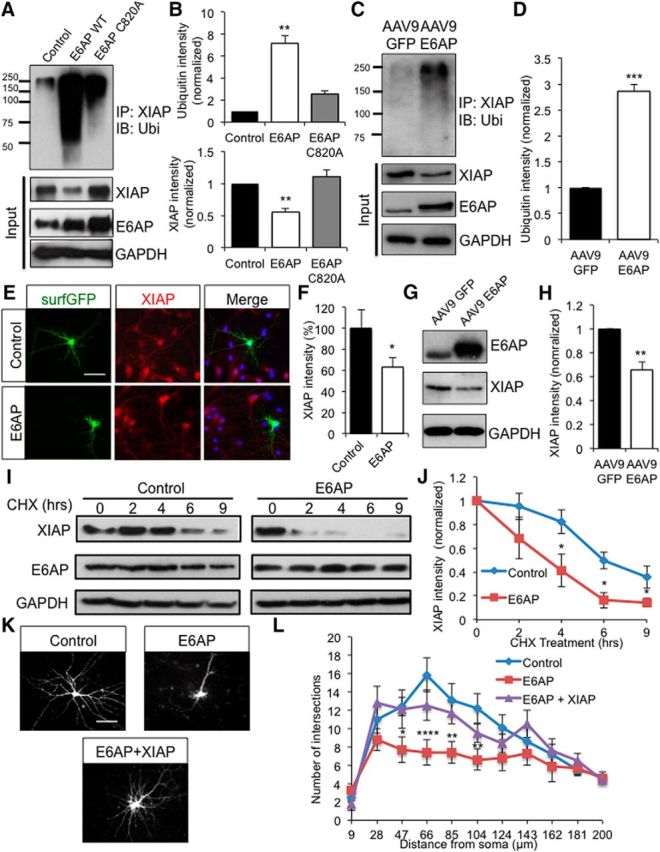
E6AP targets XIAP for ubiquitination and degradation. A, XIAP ubiquitination assay. HEK293 cells were transfected with FLAG-XIAP, HA-ubiquitin, and either a vector control, E6AP, or the E3 ligase dead mutant E6AP C820A for 2 d. XIAP was immunoprecipitated and probed for ubiquitin (ubi). Cell lysates (input) were also probed to detect total protein levels. B, Quantification of Western blot intensities. E6AP, but not E6AP C820A, caused an increase in XIAP ubiquitination and a decrease in XIAP protein levels; n = 3 independent experiments. C, D, XIAP ubiquitination assays using lysates of neurons infected with AAV9 GFP or AAV9 E6AP virus for 10 d. Increased intensity of ubiquitination signals on XIAP was detected; n = 3 independent experiments. E, Immunostaining of endogenous XIAP (red) in neurons transfected with surfGFP (green) or together with E6AP. Nuclei were indicated by DAPI staining (blue). Scale bar, 50 μm. F, Quantification of the XIAP signal intensity relative to the control; n = 10 cells per condition. G, H, Neurons were infected with AAV9 GFP virus or AAV9 E6AP virus for 10 d, and XIAP levels were measured by Western blot. Quantification showed a reduced level of XIAP in E6AP-infected neurons; n = 3 independent experiments. I, Degradation assay of XIAP with or without E6AP. Transfected HEK cells were treated without cycloheximide (CHX) for various time points and cell lysates were collected to examine XIAP levels by Western blot. J, Quantification of the degradation rate of XIAP over time; n = 4 independent experiments. K, Morphology imaging of primary neurons transfected with surfGFP alone or together with E6AP or E6AP + XIAP. The effect on dendritic arborization markedly decreased in neurons expressing E6AP C820A (Fig. 4-1, ). Scale bar, 50 μm. L, Sholl analysis showing a blockade of the E6AP effect in dendritic remodeling by XIAP overexpression; n = 10 cells per condition. Error bars represent SEM, *p < 0.05, **p < 0.01, ***p < 0.001, ****p < 0.0001.