
Figure 5.
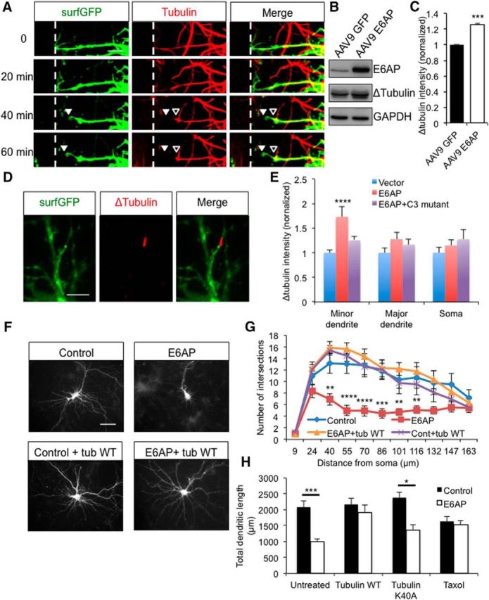
Microtubule cleavage and retraction in E6AP-induced dendritic remodeling. A, Neurons were transfected with surfGFP and pTRE-E6AP for 24 h, and loaded with SiR-Tubulin, a fluorogenic and cell-permeable dye for tubulin labeling, before being treated with doxycycline (Dox) to induce E6AP expression. Tubulin and surfGFP images were obtained every 20 min for 12 h following Dox application. Representative images show that retraction of microtubule (red; hollow arrowhead) occurred before that of the GFP-positive dendritic branch (green; solid arrowhead). The original position of the dendritic tip is indicated by a dashed line. Scale bar, 5 μm. B, Neurons were infected with AAV9 GFP virus or AAV9 E6AP virus for 10 d, and cleaved tubulin levels were measured by Western blot with an antibody specifically against the cleaved microtubule (ΔTubulin). C, Quantification showed an increased level of microtubule cleavage in E6AP-infected neurons; n = 3 independent experiments. D, Representative image of E6AP neurons immunostained with ΔTubulin. Scale bar, 10 μm. E, Quantification of ΔTubulin immunointensity in neurons transfected with vector control, E6AP, or E6AP + Casp3 C163A (E6AP + C3 mutant), compared with control; n = 10. F, Morphology of neurons transfected with surfGFP, tubulin WT, E6AP, or E6AP + tubulin WT. Scale bar, 50 μm. G, Sholl analysis of dendritic arborization; n = 10 cells per condition. H, Quantification of total dendritic length; n = 10 cells per condition. Changes in tubulin stabilization also affected the E6AP-dependent dendritic remodeling (Fig. 5-1, . Error bars represent SEM, *p < 0.05, **p < 0.01, ***p < 0.001, ****p < 0.0001.