Figure 7.

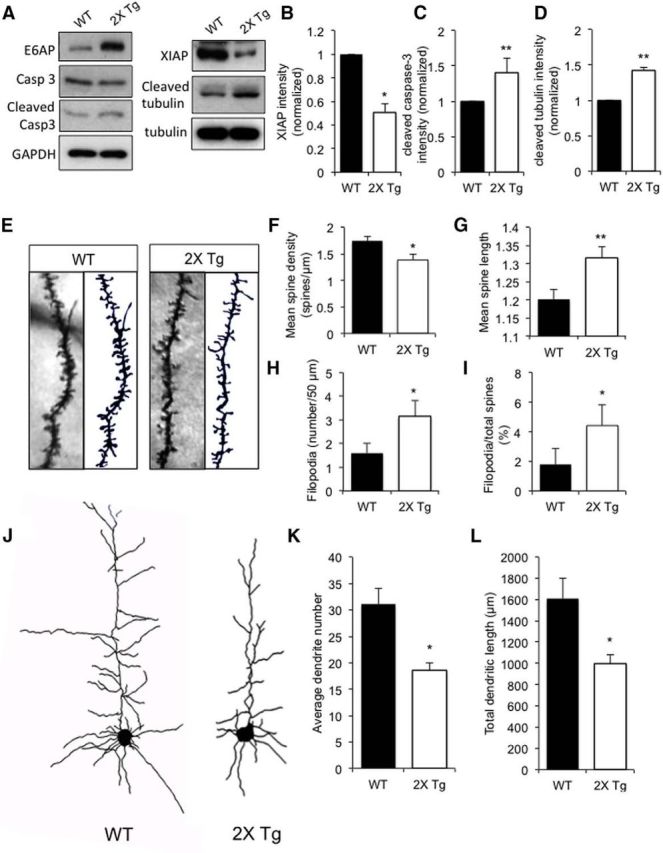
E6AP autism mouse model neurons show impairment in spine maturation and reduction in dendritic branching. A, Brain lysates collected from WT or 2X Tg mice at P15 were probed for E6AP, XIAP, caspase-3, cleaved caspase-3, cleaved tubulin, and total tubulin. GAPDH was also probed as a loading control. B–D, Quantification analysis of Western blots for XIAP, cleaved caspase-3, and cleaved tubulin; n = 3 for each. E, At P15, brains of WT and 2X Tg mice were subjected to Golgi staining. Representative images of spine morphology of layer-V somatosensory cortical neurons are shown. F, Mean spine density decreased in 2X Tg mice; n = 10 neurons. G, Mean spine length increased in 2X Tg mice; n = 10 neurons. H, I, The percentage and number of filopodia increased in 2X Tg mice; n = 10 neurons. J, Representative layer-V pyramidal neuron tracing images of Golgi staining from P15 WT and 2X Tg mouse brain slices. K, L, Measurement of average dendrite number and total dendritic length in pyramidal neurons; n = 12 neurons. Error bars represent SEM, *p < 0.05, **p < 0.01 (Fig. 7-1, ). Summary of the E6AP-dependent dendritic remodeling pathway.
