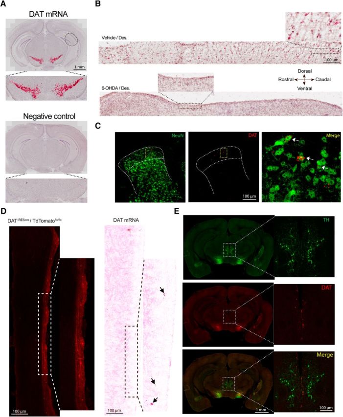
Figure 2.

Spinal DA lesion reduces the number of TH-immunoreactive neurons in A11 and spinal cord DAT expression. A, DAT in situ hybridization probe shows a specific signal in the substantia nigra, whereas the negative control probe shows no signal. B, Parasagittal sections of the spinal cord show DAT mRNA expression in the dorsal horn. Red punctate are visible throughout the rostrocaudal axis, whereas spinal DA lesion drastically decreases the level of DAT mRNA expression. N = 3 per group. C, Coronal section of the spinal cord showing DAT mRNA expression in the dorsal horn mainly localized in superficial laminae and overlapped with a neuronal marker (NeuN). N = 3 per group. D, Illustration of the DAT expression pattern in a DATIREScre/ROSA-LSL-tdTomato animal as well as DAT mRNA expression using RNAscope. E, Representative coronal section of the brain showing a high level of TH immunoreactivity in the A11 nucleus, whereas DAT mRNA signal is not readily detected in this region. Mice in these experiments were female and male.
