Figure 3.
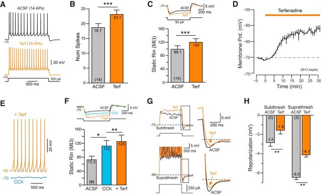
ERG-mediated responses in neocortical pyramidal cells. A, Terfenadine increases the number of spikes evoked by depolarizing current stimuli (comparison from ACSF to Terf). B, Plot of results from 14 experiments similar to A. ***p = 2.93E-04, T = 4.89, df = 13, paired t test. C, Terfenadine increases input resistance assayed from similar reference membrane potentials (approximately −70 mV). ***p = 1.50E-04, T = 5.28, df = 13, paired t test. D, Plot of mean membrane depolarization evoked by Terf in three pyramidal cells. E, Following cholinergic stimulation with CCh (blue trace), the same concentration of Terf elicited spontaneous firing (orange trace). F, Plot of input resistance assayed at −70 mV in ACSF, following CCh and with CCh + Terf. *p = 0.027, T = 3.74, df = 5; **p = 0.0058, T = 5.4253, df = 5, paired t test. G, Terfenadine attenuates poststep repolarization to both subthreshold (top) and suprathreshold (bottom) responses to depolarizing steps. Enlargements of repolarization shown on the right. H, Plot of the attenuation of the poststep repolarization. Subthreshold: **p = 0.00787, T = 4.93, df = 4; **Suprathreshold: p = 0.00167, T = 7.53, df = 4. Both paired t tests.

