
Figure 9.
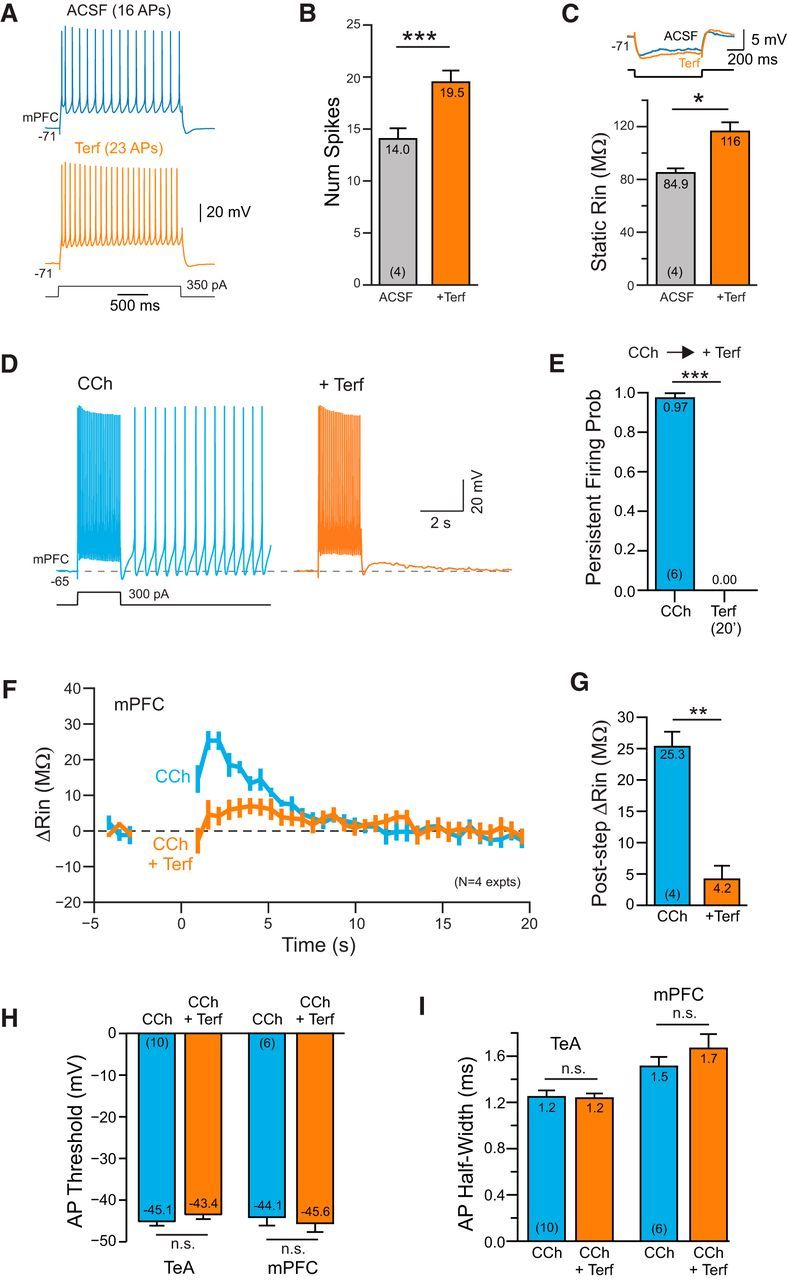
ERG-mediated persistent activity in prefrontal cortical neurons. A, Terfenadine increases the number of APs evoked by depolarizing steps in L5 mPFC pyramidal cells. B, Summary plot of number of APs evoked by the conditioning step in ACSF and Terf. *p = 0.0131, T = 5.30, df = 3, paired t test. C, Plot of the increase in input resistance with Terf. Example responses above plot. ***p = 8.52E-05, T = 29.54, df = 3, paired t test. D, Terfenadine abolishes persistent firing evoked in CCh. E, Summary plot of effectiveness of Terf in blocking persistent firing in CCh in 6 experiments. ***p = 3.62E-07, T = 34.93, df = 5, paired t test. F, Plot of the change in input resistance in 4 mPFC pyramidal cells following conditioning depolarizing steps in CCh and CCh + Terf. Experimental protocol is the same as shown in Figure 6, B and C. G, Summary plot of maximal increase in input resistance observed during ADP response. **p = 0.00163, T = 10.95, df = 3; paired t test. H, Plot of AP threshold in CCh before and after Terf. Left column pair in TeA neocortex and right pair in medial prefrontal cortex. TeA: p = 0.25; PFC: p = 0.40; paired t test. I, Plot of AP half-width calculated at the half-maximal AP amplitude. Same column order as H. TeA: p = 0.78; PFC: p = 0.33, paired t test.