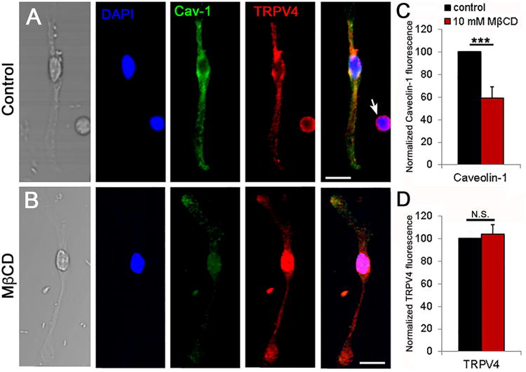
Figure 2. Cholesterol loss disassembles caveolae without impacting on the expression and trafficking of TRPV4.
(A) Representative Müller cell immunolabeled for TRPV4 (Alexa 594 nm), Cav-1 (Alexa 488 nm) and DAPI; an adjacent putative RGC shows TRPV4-ir but does not stain for Cav-1 (arrow). (B) Representative cell following 1 hour incubation in MβCD. Scale bar = 20 μm (C) Averaged summary showing that MβCD significantly reduces the fluorescence intensity of Cav-1-ir (P < 0.001, n = 4 separate experiments); (D) MβCD has little effect on the intensity of TRPV4-ir (P = 0.733).

