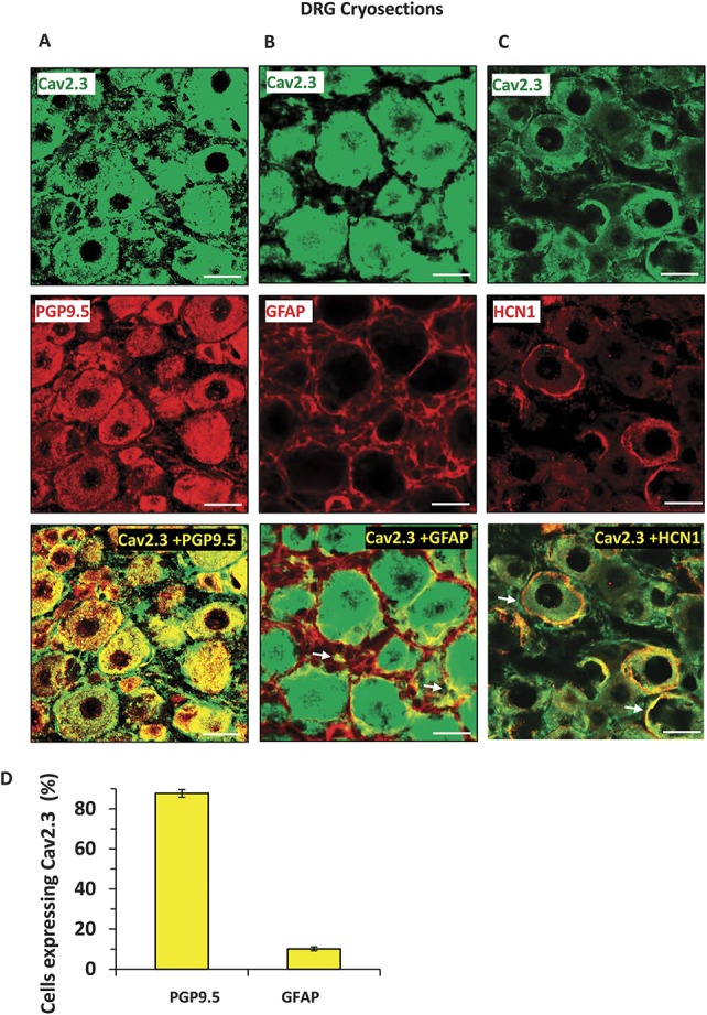
Figure 4.

Analyses of expression of the Cav2.3 protein in neuronal and nonneuronal cells or in cell membrane of the cells in dorsal root ganglia in vivo. Representative images to demonstrate expression of Cav2.3 in mouse DRG and colabeling with PGP9.5-positive neuronal cells (A), GFAP-positive satellite cells (B) and with HCN1 (hyperpolarization-activated cyclic nucleotide-gated) channel in the cell membrane (C). Observed colocalization is highlighted with white arrows. Quantification of coexpression of each neuronal subtype with the Cav2.3 expressing neurons is shown in panel D. Scale bars represent 50 µm in all panels. Tissue samples from 3 independent mice were analyzed.
