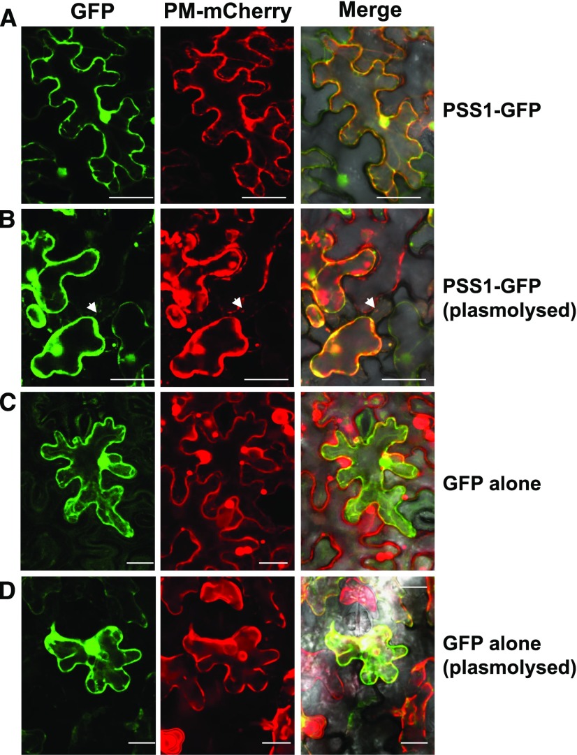
Figure 6.
PSS1 is localized to the plasma membrane. A, PSS1-GFP fusion and mCherry-tagged plasma membrane (PM) marker Arabidopsis PIP2A colocalized to plasma membrane of the epidermal cells of N. benthamiana. B, The colocalized PSS1-GFP and PIP2A-mCherry fluorescent proteins remain as a complex following plasmolysis with 1 m NaCl. C, Control GFP fluorescent protein was localized to cytoplasm. D, Plasmolysis of the cell coexpressing the GFP and PIP2A-mCherry proteins. White arrowheads indicate Hechtian strands (for details, see Supplemental Fig. S5). Bars = 50 μm for PSS1-GFP and 25 μm for GFP alone.

