Figure 2.
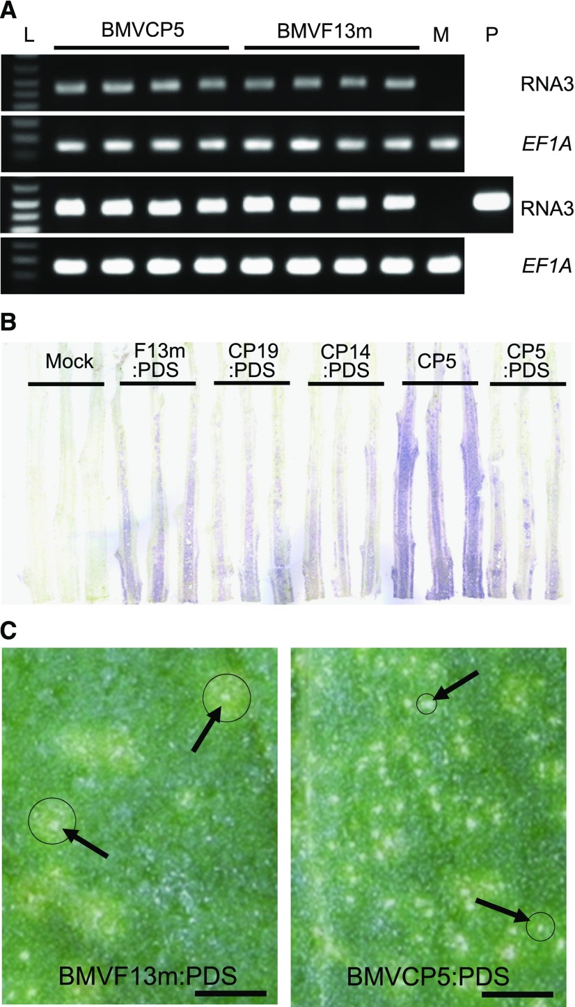
Modified BMV vectors can accumulate similarly to the original BMV vector in N. benthamiana after agroinfiltration. A, Accumulation of BMVCP5 and BMVF13m in N. benthamiana leaves at 4 dpai as measured through semiquantitative RT-PCR. PCR product from BMV genomic RNA3 after 25 cycles (top) and 30 cycles (bottom) is shown. Maize translation elongation factor 1A (ZmEF-1A) transcript levels were used as internal normalization controls. Each lane represents PCR product from an individual plant. Lane L is DNA ladder. Lane M represents amplification from extract of mock-inoculated tissue, as negative control. Lane P represents amplification from plasmid pC13/F3CP5 as positive control. B, Accumulation of BMV vectors in N. benthamiana stems at 10 dpai. BMV CP was detected through a tissue-print assay using a polyclonal antibody against the BMV CP. Purple color in stem longitudinal prints indicates positive detection of the virus CP. The experiment was repeated twice with similar results. C, Local lesions (noted by arrows) imaged at 6 dpi with BMVF13m:PDS or BMVCP5:PDS in leaves of C. amaranticolor. Strong chlorotic rings outside of necrotic lesions are marked with circles. Bars = 7 mm.

