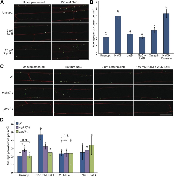
Figure 6.
Salt-induced peroxisome proliferation depends on actin polymerization. A, Latrunculin B blocks salt-induced peroxisome division. Here, 3-d-old Col-0 GFP-PTS1 seedlings grown on unsupplemented media at 22°C under continuous light were transferred to the indicated treatments for 16 h. Seedlings were then counterstained with propidium iodide before imaging by confocal microscopy. Signal from GFP-PTS1 was false-colored green and signal from propidium iodide was false-colored red. Scale bar = 25 μm. B, Mean number of peroxisomes (± se) from 3-d-old Col-0 GFP-PTS1 seedlings treated as indicated. Peroxisome numbers in seedlings treated with oryzalin + NaCl are statistically indistinguishable from seedlings treated with NaCl alone (using ANOVA), whereas peroxisome numbers on latrunculin + NaCl are statistically indistinguishable from peroxisome numbers in seedlings grown on unsupplemented media and latrunculin alone (using ANOVA). C, Confocal images of 3-d-old wild type (Col-0), mpk17-1, and pmd1-1 carrying the 35S:GFP-PTS1 reporter (Zolman and Bartel, 2004) treated for 16 h as indicated. Scale bar = 25 μm. Seedlings were counterstained with propidium iodide before imaging; signal from GFP-PTS1 was false-colored green and signal from propidium iodide was false-colored red. D, Mean number of peroxisomes (μ se) from 3-d-old wild type (Col-0), mpk17-1, and pmd1-1 carrying the 35S:GFP-PTS1 reporter (Zolman and Bartel, 2004) treated for 16 h as indicated. Peroxisome numbers in mpk17-1 and pmd1-1 do not significantly change with the addition of NaCl or with latrunculin + NaCl. Data analyzed using ANOVA and Tukey’s HSD (letters) and by a two-tailed unpaired t test (* = P ≤ 0.05). Wt, wild type.

