Figure 4.
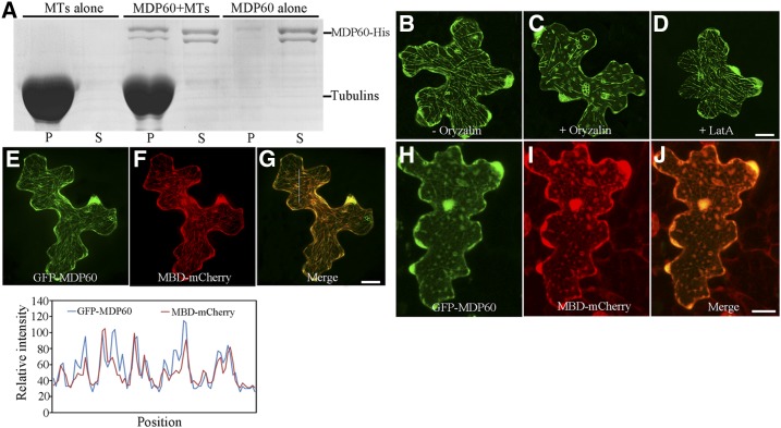
MDP60 directly binds to microtubules. A, MDP60-His was cosedimented with paclitaxel-stabilized microtubules. MDP60-His was most abundant in the supernatant (S) in the absence of microtubules, but cosedimented with microtubules into pellets (P). B, GFP-MDP60 was transiently expressed in Arabidopsis pavement cells where it formed filamentous structures. The filamentous pattern of GFP-MDP60 was disrupted when cells were treated with 10 μm oryzalin for 30 min (C) but was unaffected when treated with 100 nm LatA for 30 min (D). E to G, Colocalization of transiently expressed GFP-MDP60 and MBD-mCherry. Plot of a line scan drawn in G showing a strong correlation between spatial localization of GFP-MDP60 and MBD-mCherry. H to J, The localization could be disrupted via oryzalin. Bars in D, G, and J = 20 μm.

