Figure 1.
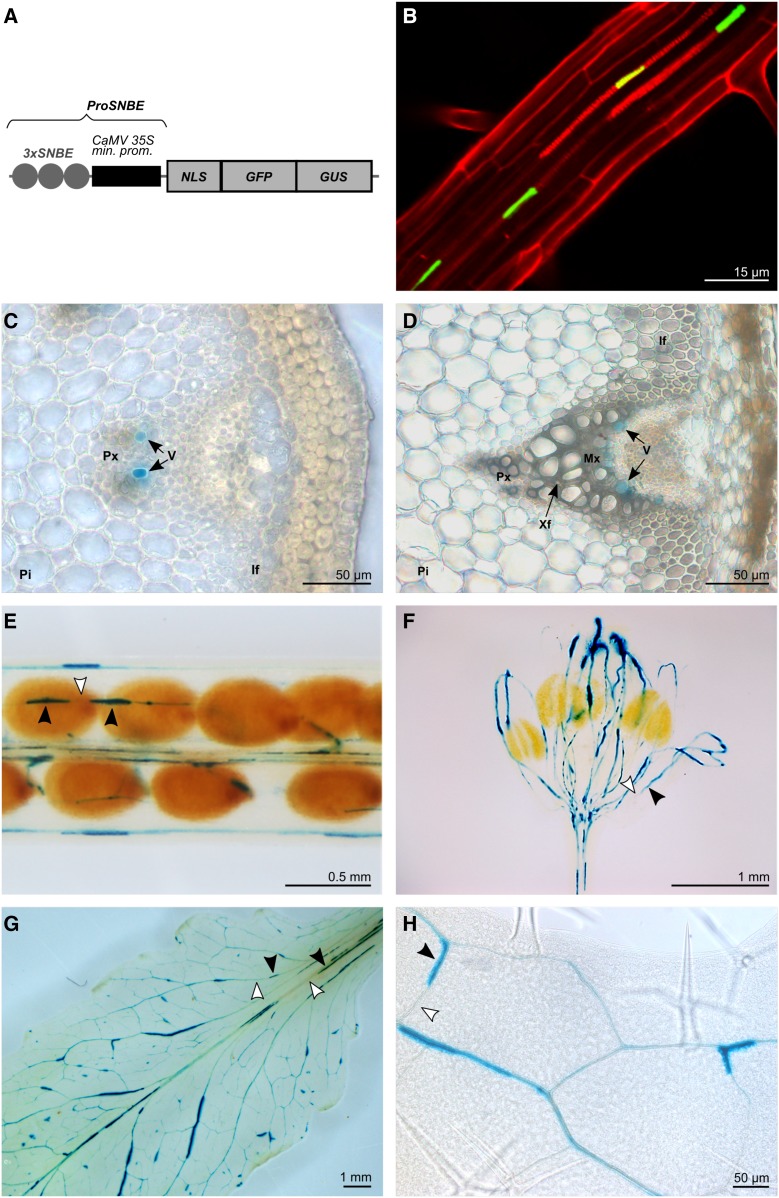
Expression pattern conferred by ProSNBE. A, Diagram of the ProSNBE:GFP:GUS construct: GFP and GUS reporter genes are driven by three copies of the XCP1-SNBE sequence fused to the CaMV minimal 35S promoter (ProSNBE). NLS, Nuclear localization signal. B, Expression analysis in roots showing GFP in xylary vessels. Propidium iodide was used to counterstain the cell wall. C, Cross section of an elongating internode of the primary inflorescence stem showing GUS staining in developing vessels of the protoxylem. D, Cross section of a nonelongating internode of the primary inflorescence stem showing GUS staining in developing vessels of the metaxylem but not in xylary or interfascicular fibers. E to H, GUS expression analysis in siliques (E), flowers (F), and rosette leaves (G and H), showing reporter gene expression in the vasculature. Black arrowheads indicate cells with GUS staining, and white arrowheads indicate cells lacking GUS staining. For B, transgenic ProSNBE:GFP:GUS seedlings were grown for 20 d in a long-day photoperiod. For C to H, transgenic ProSNBE:GFP:GUS plants were grown for 6 weeks in a short-day photoperiod followed by 5 weeks in a long-day photoperiod. If, Interfascicular fibers; Mx, metaxylem; Pi, pith; Px, protoxylem; V, vessel; Xf, xylary fiber.

