Figure 4.
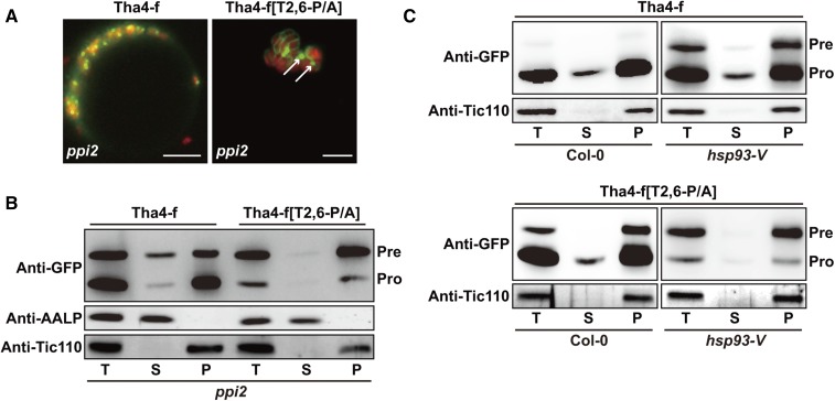
TPs lacking Pro residues cause mistargeting of TMD-containing preproteins to outer envelope membranes. A, Localization of reporter proteins. The ppi2 protoplasts were transformed with the indicated constructs, and GFP patterns were observed 12 h after transformation. Green, red, and yellow signals represent GFP, chlorophyll autofluorescence, and the overlap between green and red signals, respectively. Scale bar = 20 μm. B and C, Subcellular fractionation. Protein extracts were separated into soluble and pellet fractions by ultracentrifugation, and fractions were analyzed by western blotting with anti-GFP antibody. AALP and Tic110 were used as controls for soluble and membrane proteins, respectively. T, total protein; S, soluble fraction; P, pellet fraction; Pre, precursor form; Pro, processed form.

