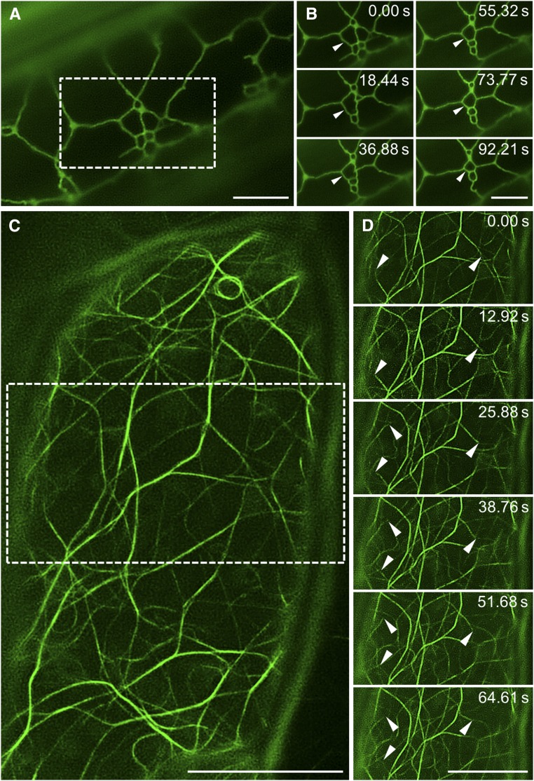
Figure 4.
The potential for time-resolved structured illumination microscopy imaging. A and B, Overview (A) and time-lapse imaging (B) of cortical endoplasmic reticulum in a hypocotyl epidermal cell expressing a GFP-HDEL marker. The dynamic reorganization of the endoplasmic reticulum tubular structure is depicted by arrowheads (B). C and D, Overview (C) and time-lapse imaging (D) of actin filaments in a hypocotyl epidermal cell expressing a GFP-FABD2 marker. The dynamic reorganization of actin filaments is depicted by arrowheads (D). Bars = 5 μm (A and B) and 10 μm (C and D).

