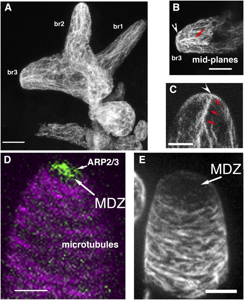
Figure 2.
Actin and microtubule organization in Arabidopsis leaf hairs and cotton fibers during the process of cell elongation and tapering. A, Whole mounted trichome with branches that are becoming progressively tapered (defined as stage 4). The cell is labeled with an anti-actin antibody using the freeze-shattering technique. Note prominent tip actin in branches 2 (br2) and 3 (br3). B, Midplanes of br3 in A showing cortical actin at the apex and cytoplasmic bundles that are oriented toward the apical meshwork. C, Whole mounted trichome labeled with phalloidin. Apical actin meshwork and cytoplasmic bundles are labeled as in B. D, Live-cell image of GFP-tagged ARP2/3 complex (green) and microtubules (magenta). ARP2/3 localizes within the apical microtubule-depletion zone (MDZ) and is required to polymerize the apical actin meshwork. E, Whole mounted 1-DPA cotton fiber in the process of cell tapering labeled with an anti-α-Tubulin antibody. Bars = 5 μm.

