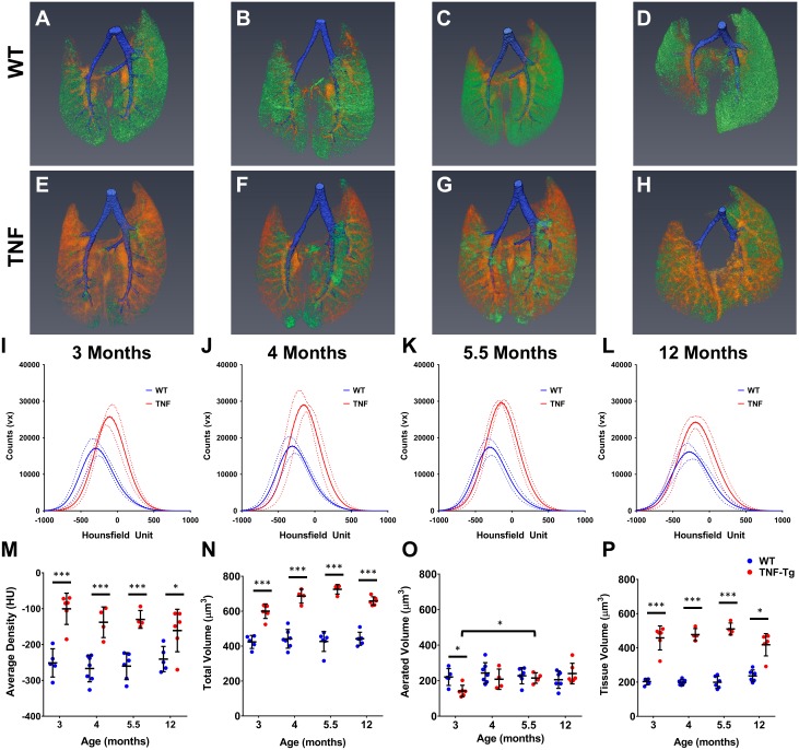
Fig 3. TNF-Tg male mice have increased tissue volume compared to WT littermates measured via μCT.
Representative 3D reconstructions of 3 (A, E), 4 (B, F), 5.5 (C, G), and 12 (D, H) month old WT and TNF-Tg male mice show a clear increase in tissue volume (Orange-Red) in the TNF-Tg animals at all timepoints while maintaining a similar amount of aerated volume (Green) at all timepoints. Conducting airways that were segmented out of analysis shown in Blue. Histograms of the extracted data from the whole lung segmentation for each timepoint is presented in I-L (M ± 95%CI, n = 4–6). TNF-Tg male mice have a statistically significant increase in mean lung intensity (M), total lung volume (N) and tissue lung volume (P) at all timepoints compared WT littermates (*p<0.05, ***p<0.001, M±SD, n = 4–6).

