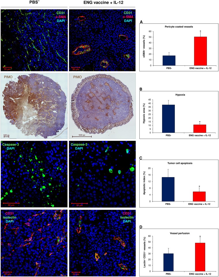
Fig 9. Effect of combined therapy (ENG vaccine + IL-12) on tumor blood vessels.
On 20th day of the combined therapy, mice were sacrificed and tumors were excised for immunohistochemical staining. “Normalized” vessels were identified using several tests [35]: (A) α-SMA and CD31 staining was used to identify pericytes-covered tumor vessels (α-SMA+CD31+ vessels, percentage of CD31+ vessels; n = 6; 10 visual fields per tumor section; magnification 20×; *P <0.001, the Cochran’s C test). (B) Staining with pimonidazole (PIMO) was conducted to visualize hypoxic regions in tumors (PIMO+ area (% of tumor area); n = 5; magnification 4×; *P <0.001, the Student’s test). (C) Caspase-3 staining was used to identify cancer cells that undergo apoptosis during vasculature “normalization” (apoptotic index: caspase-3+/ total cells; n = 6; 10 visual fields per tumor section; magnification 40×; *P <0.001, the Mann-Whitney U test). (D) Lectin perfusion test was used to assess vessel permeability in tumors (lectin+CD31+ vessels (% of CD31+ vessels); n = 6; magnification 20×; *P <0.001, the U Manna-Whitneya test). The structure of tumor vessels in mice treated with combined therapy resembles a regular one: with open lumens, the walls are thick, with a fine layer of pericytes (αSMA) adjacent to their surface (A). Smaller areas of hypoxia (B) and lower number of cells undergoing apoptosis were also found in tumors of treated mice (C). Increased number of perfused blood vessels (lectin+CD31+) were observed in tumor sections from mice treated with combined therapy (D). This indicated that the B16-F10 tumor vasculature in ENG vaccine with IL-12- treated mice is mature and functional.

