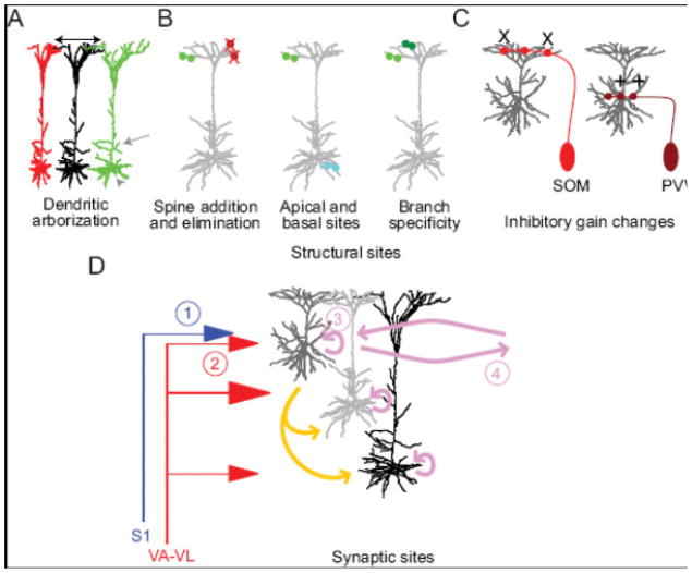
Figure 4. Structural and synaptic sites of plasticity in M1 during motor skill learning.
A range of sites for cortical plasticity have been examined. Structural sites include (A) changes in the size of the dendritic arbor (black arrows) of pyramidal neurons in the topographic region where plasticity occurs (Greenough et al., 1985; Withers and Greenough, 1989). Gray arrow and arrowhead in (A) indicate addition and elimination of dendritic surface possible during plasticity. (B) Changes in spine addition (green and cyan), elimination (red X), and stabilization (Xu et al., 2009; Yang et al., 2009) occur, including differences in apical and basal locations (Peters et al., 2014) and branch specificity or clustering (dark green versus light green; Fu et al., 2012; Yang et al., 2014). (C) Structural changes in interneuron axons, including addition (+) or loss (X) in an interneuron subtype-specific manner (Chen et al., 2015) are also reported. (D) We illustrate here the putative changes in local, horizontal, and long-range inputs as a result of motor skill learning. Long-range inputs that may change include (1) S1 inputs to L2/3 neurons and (2) VL inputs to L2/3 neurons (Baranyi and Feher, 1981; Iriki et al., 1989; Kaneko et al., 1994; Sakamoto et al., 1987), albeit with distinct learning rules for corticocortical and thalamocortical plasticity. Changes within motor cortex include L2/3 (3) local (Aroniadou and Keller, 1995) and (4) long-range horizontal connections (Hess et al., 1996; Hess and Donoghue, 1994; Rioult-Pedotti et al., 2000; Rioult-Pedotti et al., 1998). Other intralaminar (pink) and translaminar (gold) local connections are also putative sites of synaptic changes.

