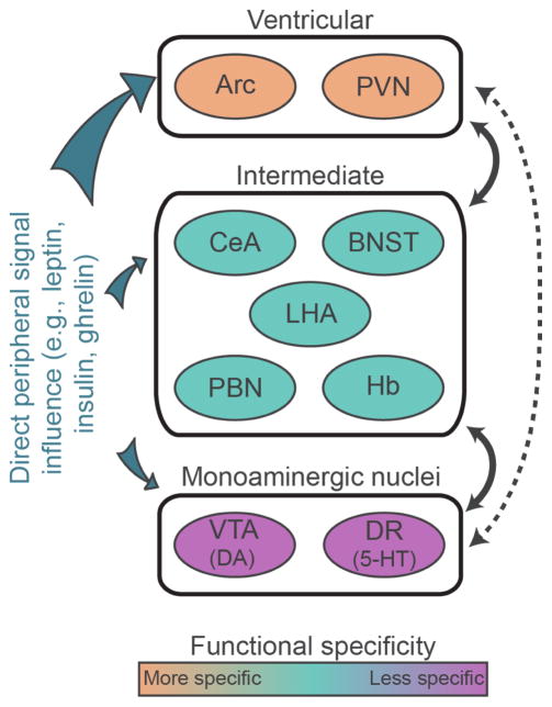
Figure 1.
Circuits involved in feeding and reward. Schematic illustration of ventricular, intermediate, and monoaminergic nuclei. There are strong reciprocal connections between ventricular and intermediate as well as intermediate and monoaminergic nuclei (indicated by solid lines). Direct connections between ventricular neurons and monoaminergic nuclei are relatively sparse or absent in adults (indicated by the dashed line). Ventricular neurons express receptors for peripheral signaling molecules implicated in feeding are more densely than do intermediate and monoaminergic nuclei. Arc, arcuate nucleus; BNST, bed nucleus of the stria terminalis; CeA, central nucleus of the amygdala; DA, dopamine; DR, dorsal raphe; Hb, habenula; LHA, lateral hypothalamic area; NAc, nucleus accumbens; PBN, parabrachial nucleus; PFC, prefrontal cortex; PVN, paraventricular nucleus of the hypothalamus; VTA, ventral tegmental area; 5-HT, 5-hydroxytryptamine.

