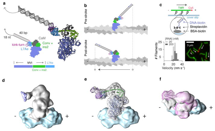
Figure 1. Design and characterization of an engineered myosin with an RNA lever arm.
a, Design of protein and RNA components. An annotated schematic is shown alongside a larger 3-dimensional ribbon diagram for M6-RB:ktL. Below: protein block diagram for M6-RB. The RNA-binding L7Ae domain is fused to myosin VI (MVI) after the converter domain and insert 2 (conv + ins2). b, Cartoon of predicted power stroke for M6-RB:ktL bound to actin. In the transition from the pre-stroke to the post-stroke state, the tip of the lever arm moves toward the minus end of the actin filament. c, Measuring directed motility using a gliding filament assay. Top: assay design. M6-RB:ktL is affixed to the surface by binding to a complementary biotinylated DNA strand immobilized via streptavidin and biotin-BSA. Propelled actin is fluorescently labeled with Cy5 at its plus end, and TMR along its body. Bottom: results. Image is taken from a movie of gliding filaments, with arrows showing direction of motion. A stacked histogram of filament velocities is shown for two surface [RNA] conditions (see also Supplementary Table 1 and Supplementary Movies 1a–b). d–f, Cryoelectron microscopy reconstructions of engineered myosin and RNA bound to actin, low-pass filtered at 13 Å. d, Reconstruction of M6-RB (apo, grey) bound to F-actin (blue). Segmented density is displayed corresponding to a single myosin motor bound to 2 actin subunits. A difference map of M6-RB:ktLshort (apo) minus M6-RB (apo) is displayed as a purple isosurface, unambiguously localizing the position of the RNA. e, Flexible fitting of myosin-RNA model (Supplementary Movies 3b,c) to the cryo-EM reconstruction of M6-RB:ktLshort (apo) bound to F-actin. The DireX flexible-fitting model of M6-RB:ktLshort (apo) is colored as in Fig. 1a. f, Nucleotide-dependent conformational change (Supplementary Movies 4a,b). Overlaid reconstructions were segmented as in Fig. 1e for the apo (grey surface) and ADP (pink mesh) nucleotide states of M6-RB:ktLshort bound to actin (blue surface) .

