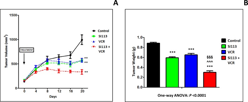
Figure 6. SI113 synergizes with VCR in restraining the growth of ADF xenografts in immunocompromised mice.
ADF cells (2.5 × 10⁶ cells) were implanted in the flanks of NOD/SCID female mice. When tumors reached the volume of about 130 mm3, mice were divided into four arms, i.e. Control, SI113, VCR or SI113 plus VCR for the in vivo treatment. Drugs (or their solvents in the Control arm) were administered five days/week, and tumor growth was monitored every 4 days for 20 days after the beginning of the treatment. (A) The graph shows the curves for tumor growth for control and animals treated with SI113, VCR or their association. Statistical analysis was done using the Student’s two-tailed t test (** = P < 0.01). Data are expressed in mm3 ± Standard Error (SE). (B) At day 20 from the beginning of the treatment, mice were sacrificed and tumors were excised and weighed. The histogram represents tumor weight for each experimental arm expressed in g ± SE. In this panel, statistical analysis among groups was done using the One-way ANOVA test followed by the Tukey’s Multiple Comparison Test. Statistical significance is also indicated (*significance vs. Control; ^significance vs. SI113; §significance vs. VCR).

