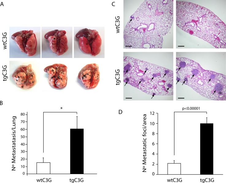
Figure 6. Platelet C3G favors melanoma metastasis.
TgC3G and wtC3G mice were injected with melanoma B16-F10 cells. Lungs were removed 15 days after tumor cell injection, and surface metastases were quantitated. (A) Macroscopic pictures (three representative examples from each of the groups) showing a clear increase in the number of metastatic foci in tgC3G lungs, as compared to lungs from control mice. (B) Surface metastases per lung were counted in both groups. The graph shows the average number of metastases in each group (n = 4 mice per genotype). *p < 0.05. (C) Representative lung sections of two mice from each genotype, showing hematoxylin/eosin staining. Arrows point to metastatic foci in the lung sections. Scale bars: 10 µm. (D) Values in the graph represent the number of tumor foci per area (n = 12 lung sections per group analyzed). P-value comparing the groups is shown.

