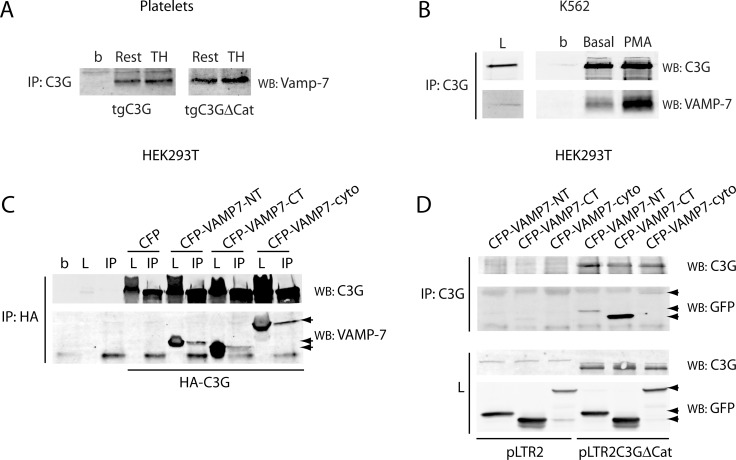
Figure 7. C3G interacts with Vamp-7 in platelets from tgC3G and tgC3G∆Cat mice.
(A) Immunoprecipitation of C3G from resting (Rest) and thrombin (TH, at 0.2 U/mL) stimulated platelets for 5 min at 37°C under stirring. Vamp-7 in the immunocomplexes was detected by Western blot with anti-TI-VAMP antibody (Santa Cruz Biotechnologies, sc-67060). Platelet cell lysate from 4 mice of each genotype was used. b: lysate incubated with agarose beads. (B) Immunoprecipitation of C3G from stably C3G-overexpressing K562 cells untreated (basal) or treated with 20 nM PMA for 10 min. C3G and VAMP-7 in the immunocomplexes were detected with anti-TI-VAMP and anti-C3G antibodies. L: total cell lysate (50 µg). b: lysate incubated with agarose beads. (C) HEK293T cells were transiently transfected with a HA-tagged C3G construct together with the indicated CFP-VAMP-7 fusion proteins or the empty pCDNA3-CFP-C4 plasmid (CFP). Ectopic C3G was immunoprecipitated with anti-HA.11 monoclonal antibody and C3G and VAMP-7 detected with anti-C3G and anti-TI-VAMP (arrows). b: agarose beads incubated with non-transfected lysate. L: total cell lysate (50 µg). IP: immunoprecipitated with anti-HA.11. (D) HEK293T cells were transiently transfected with pLTR2C3G∆Cat construct or the empty pLTR2 vector [33], together with the above CFP-VAMP-7 constructs. C3G was immunoprecipitated with anti-C3G antibodies (IP) and C3G and VAMP-7 detected with anti-C3G and anti-GFP (Santa Cruz Biotechnologies, sc-9996) antibodies. L: total cell lysate. VAMP7-NT: N-terminal (longin) domain of VAMP-7 (aminoacids 1-120); VAMP7-CT: C-terminal (SNARE) domain of VAMP-7 (aminoacids 121-188); VAMP7-cyto: VAMP-7 cytosolic domain (aminoacids 1-188).

